Wednesday, January 8, 2014

An insect bibliophile


I found this little insect, which is less than  two millimetres long, in a bag of walnuts imported from France. It's a psocid, commonly known as a booklouse, on account of the fact that these often turn up in the bindings of old books that have been stored in a damp place. They also like wallpaper and we used to find them behind peeling damp wallpaper in our house, before we stripped it all off and redecorated, thereby making scores of booklice homeless; I suspect they feed on wallpaper paste. 

Psocids are also found on tree bark and are sometimes known as barklice.


Psocids look superficially like very small aphids but instead of a piecing feeding stylet they have biting jaws, that are visible in this side view.


Some species are wingless, others have wings that they fold over their backs like a tent, but this one only has vestigial wings - or perhaps they are wings that have yet to develop fully.


Other distinctive features are the relatively large compound eyes positioned on either side of the head and the long antennae.

Saturday, October 26, 2013

Overheating sweet potato?


One of the best ways to learn more about food is to grow some of the fruit and vegetables from exotic fresh produce that you can buy in supermarkets. Over the years I've grown avocados, litchi, rice, lentils, chickpeas, figs, citrus fruits, lemon grass, ginger, chilli, cape gooseberries, squashes, passionfruit, sesame and pomegranates from supermarket produce - but this was the first time I'd ever grown sweet potatoes Ipomoea batatas.

After a slow start the tuber produces some healthy vigorous shoots but then ....


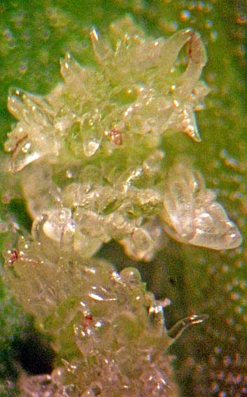
...... these white granules appeared on the leaves. At first I thought they might be some small insect pest but ...


.... a closer look revealed that they were part of the leaf surface ....and under the microscope ...


..... they turned out to be clusters of swollen, glassy secretory hairs, known botanically as colleters. They're present on upper and lower leaf surfaces.


At higher magnification you can see how inflated the hairs are. They are full of what appears to be ....


...... mucilage that flows out when they burst. 

The plant is in a flower pot on a sunny widowsill so the growth conditions are unnatural. Some older leaves have a large number of colleters, others have none. 

I don't have any clear idea of what triggers their formation or what their function would be. Some plants dump waste products in leaf surface structures like this and I wondered if they might contain calcium oxalate crystals, that are usually compartmentalised waste products in plant cell vacuoles, because sweet potato tubers do contain calcium oxalate - but I can't see any under the microscope.

Curious. I can only find two relevant research papers. 

One, from Brazil and published last year (1), describes secretory colleters in Ipomoea asarifolia, a weedy species that poisons cattle but is also used in herbal remedies.

The other, published in the Australian Journal of Botany a couple of years ago, describes similar structures on the calyx in flowers and fruit of a Ipomoea cairica (2), and the authors speculate that the secretion, which crystallises but is hygroscopic and becomes a gel in moist conditions, may have a role to play in protecting the plant from drought. That seems plausible, because the south-facing windowsill where the plant stands gets very warm on sunny days. 

So maybe by producing these colleters the plant is telling me that its too hot and needs to be watered more often ...

Sources:
1. Martins, Fabiano M.; Lima, Jamile F.; Mascarenhas, Ana Angelica S.; et al.(2012) Secretory structures of Ipomoea asarifolia: anatomy and histochemistry. Revista Brasileira de Farmacognosia – Brazilian Journal of Pharmacognosy.  Volume: 22   Issue: 1   Pages: 13-20   


2. Sousa Paiva, Elder Antonio; Martins, Luiza Coutinho (2011) Calycinal trichomes in Ipomoea cairica (Convolvulaceae): ontogenesis, structure and functional aspects. Australian Journal of Botany. Volume: 59   Issue: 1   Pages: 91-98   

Sunday, October 6, 2013

A tiny aquatic worm that clones itself

All summer, small containers of various kinds in our garden have collected rainwater and detritus - and each of these microcosms has developed a fauna of its own. This is a little oligochaete worm called Aeolosoma that I found in the layer of mouldering leaves at the bottom of one of these little pools.


Oligochaetes are annelids (segmented worms) whose bodies have only a few bristles on each segment. This species, less than two millimetres long, is almost completely transparent and has distinctive little orange spots just under the body surface. If you look closely you can also just make out the fine bristles at the junctions of the body segments. It whisks food into its mouth, which is under and towards the back of that spade-like structure called the prostomium, with fine cilia that beat and generate a water current. It's thought that that spade-like prostomium can attach like a sucker to a substrate so that the beating cilia generate suction, aiding feeding.


Aeolosoma divides asexually, budding off new individuals from the tail end, so it's quite possible that the thriving colony (I found six in a single drop of water so there must be thousands in the container) are all descended from a single original colonist.


In the image above you can see the prostomium being used rather like a vacuum cleaner nozzle.


Sunday, March 17, 2013

Fatal Attraction


Fungus gnats that emerge in swarms from soil in plant pots have become the bane of many gardeners' lives. If you grow plants in commercial potting composts on your house window ledge or in a greenhouse or conservatory, it's inevitable that you'll encounter these irritating pests because it seems that all currently available bags of potting compost are infested with them.

These little insects are scientifically known as Bradysia paupera and belong to a group known as sciarid flies. Each female can lay around 200 eggs which hatch into a worm-like, transparent larva that feeds on organic matter in the soil and also on young plant roots. A heavy infestation is capable of killing seedlings. They breed all-year-round, with overlapping generations that take less than a month to progress from egg to adult, so combating them is a constant challenge, but fortunately they have a fatal weakness - the colour yellow. They are attracted to these sticky yellow sheets of plastic that you can buy in garden centres and are glued to them as soon as their feet touch the surface.

Yellow strips of sticky plastic plastered with dead flies are unsightly but there is a more aesthetically attractive alternative - the carnivorous butterwort, Pinguicula sp., whose sticky leaves are like natural flypaper and which produces attractive flowers throughout the year. To see how effective this is, scroll down to the bottom of this post, and to see how it works, click here.


The little club-shaped structures on either side of the insect are halteres - balancing organs which smooth its flight path as its wings beat up and down.


Long-legged sciarid flies spend much of their time running around over the surface of the soil, where they lay their eggs.


Whiteflies caught on the sticky hairs of a butterwort leaf


Sciarid flies trapped on the sticky surface of a butterwort leaf


Sunday, January 6, 2013

Scale insects ....


 I recently discovered that the orchid on my desk is infested with these tiny scale insects Coccus hesperidium. Each one looks like a miniature tortoise, about 3-4mm. long and tightly attached to leaves.


The tell-tale symptom was the sticky secretion on my desk under the leaves - scale insects suck sap, like aphids, and excrete honeydew.


When I took a close look it was easy to see how the infestation had built up so rapidly. Under the heat of the microscope lamp these infants, known as 'crawlers', emerged from under their mother. The females reproduce parthenogenetically, producing about 1000 nymphs during their three month life span and sheltering the young under their shield.


When you flip a scale insect over you can see the hollow cavity which acts as a nursery for the nymphs. Each is less than 0.25mm long


Nymphs are flattened and are very active, but as they begin to develop their broad shield they settle in one spot to feed.


Here's an older individual, where you can see the shield beginning to grow outwards around the insect. The waxy shield makes these insects impervious to most insecticide sprays - so they are difficult to control. Suffocating them under horticultural oil sprays or picking them off laboriously with a paintbrush dipped in alcohol are really the only effective treatments for infected plants.


This nymph, which is about 3mm. long, has settled to feed. It's probably about a month old.


After two months they reach maturity and begin to reproduce, and then their shield grows darker as they age.

Scale insects infest a very wide range of plant hosts. You can read more about them by clicking here.


Wednesday, October 24, 2012

Mosquito larvae...


Our unusually wet summer has provided plenty of opportunities for breeding mosquitoes, with water butts and containers retaining pools of water all through the year. The water butt attached to our greenhouse has been swarming with culicine mosquito larvae.


The larvae hang from the surface film, breathing through a  siphon tube, which you can see in this image on the tail of the larva; you can see a small bubble of air attached to the siphon tip. The tiniest disturbance of the surface film sends the larvae wriggling down into the depths.


For images of an adult culicine mosquito, click here and here.


Thursday, August 30, 2012

How to Recruit an Army


Plants primarily secrete nectar as an energy source to tempt pollinators to visit their flowers, but the secretion of this substance appears to have evolved long before flowering plants appeared. Many plants, including some ferns, secrete nectar from extrafloral nectaries - i.e. nectaries in other positions on the surface of the plant. 

Legumes, like the common vetch Vicia sativa in the image above, have extrafloral nectaries on their stipules (the small, leaf-like projections on either side of the base of a leaf stalk). The extrafloral nectary is the black spot on the image above and a closer look ....


... reveals what its function might be. Ants are famous for their attraction to sweet substances and regularly visit the plant for the sugar that leaks out of these locations. This might deliver two kinds of selective advantage to the plant that would outweigh the cost of using some of its assimilated sucrose in this way. In some plants it might deflect ants, which are usually very inefficient pollinators, away from the larger source of nectar that's there to service more efficient pollinators, like bees. In other plants it may be a way of recruiting  a defensive army of ants because they become aggressive towards herbivorous insects that might try to plunder their food supply; in Acacia trees for example, the defensive benefits of hosting ants are well documented.


Extrafloral nectaries are found in a wide variety of plants and are often located on leaf petioles and mid-ribs. This is a vertical section through an extrafloral nectary on the underside of the mid-rib of a cotton plant (Gossypium sp.), stained with fluorescent dyes. The bright yellow cells at the top are xylem vessels, conducting water to the leaf blade. The very small, brick shaped blue cells below are dividing cambial cells and also phloem sieve elements that are conducting assimilated sucrose away from the leaf blade. Below that are some larger, blue-stained parenchymatous cells and then, at the very bottom, there are thin-walled finger-shaped cells which constitute the extrafloral nectary tissue, on the lower surface of the leaf mid-rib.

The blue staining is due to cellulose in the cell walls binding to a dye called calcofluor, which then fluorescence blue in UV light. You can see from this image that there's a very thin cellulose cell wall in those finger-shaped extrafloral nectary cells, because they barely fluoresce. So they easily leak sucrose that accumulates in them. The other interesting feature of this section is the orange staining in the small cells immediately above those extra-floral nectary cells. This is the endoplasmic reticulum/ Golgi complex inside the cells - the membranes and secretory vesicles that manufacture substances and transport them between cells via channels in the cell walls called plasmodesmata; these brightly-fluorescing cells seem to be highly metabolically active, so maybe the nectary cells are secreting something else, as well as sucrose.

There are some scientific papers on cotton extrafloral nectaries, their role and how they might be exploited in biological control programmes in this crop here, here and here.


Wednesday, August 8, 2012

Millions of Mites


By the time that summer arrives the foliage of most trees shows signs of insect attack, but these little eruptions on the surface of an alder leaf are caused by eriophyid mites, which are not insects but are related to spiders. I think the mite species that has produced these is Eriophyes laevis inangularis.


Each of these little domes is a chamber that's formed when the mites feed on cells on the undersurface of the leaf, leading to uneven growth that results in the formation of  a pouch where the mites can feed and breed.


This is the underside of the leaf, with the little yellow, sausage-shaped mites crawling around the entrances to the chambers, which are lined with nutritive cells that provide sustenance for the mites.


Here they are at higher magnification .........


............ and at still higher magnification, when the elongated body with four legs at the head end is visible in the mite in the top, left-hand corner. Each chamber is home to a brood of mites and a tree with a severe infestation could be covered with hundreds of thousands of them. Eriophyid mites also commonly infest sycamore and field maple leaves, producing large numbers of red pouches on the leaf surface.


These are three of the mites, each being about one fifth of a millimetre long, with only four legs.


The outer cuticle of the animal has a distinct pattern that differs between species, although the easiest way to identify species is via the symptoms that they cause on the host plant.


Here is the head, legs and cuticle patterning at higher magnification.


In addition to infesting sycamore, field maple and alder leaves eriophyid mites also attack many other plants, including goosegrass (aka cleavers) Galium aparine, whose growth is distorted by Eriophyes galii.


Typically, infested leaves curve inwards at the edges and become spoon-shaped, like the bottom, second-from-the-left leaf in this picture.


Here's the goosegrass eriophyid - the dark, globular structure top left is an air bubble on the microscope slide.


In this view you can see some of the surface patterning and an internal structure - perhaps an egg?- 


... and in this plane of focus the surface pattern of the cuticle is apparent.

Monday, July 16, 2012

Aphids in a Savage Landscape


When aphids infest plants they tend to find a good spot to feed and then stay in one place, where they'll insert their stylets into the plant's phloem, tap its sugary sap and then settle down to reproduce


When you take a close look at plant surfaces you can sometimes see why these pests are more or less sedentary. Many plants, like this goosegrass Galium aparine, are covered with epidermal hairs (trichomes) that make it difficult to tiny aphids to move around.


In the case of goosegrass the hooked hairs are primarily for attaching their weak stems to supports as they grow, but those curved spines are also awkward obstacles for minute aphids to negotiate.